Healthcare
Generate images like this by single click. Just edit the text and regenerate the image to get a new one.
Generate images for healthcare projects.

A vibrant illustration of a heart for a cardiovascular medication packaging, using a palette of red, blue, and white with soft gradients to depict a healthy, pulsating heart. The background should be a soft blue to symbolize tranquility and trust, with a photorealistic quality to the heart.
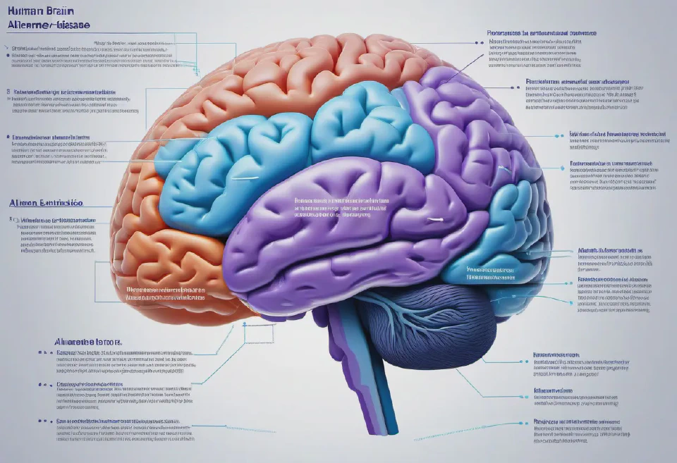
Diagram of the human brain, with areas affected by Alzheimer’s disease highlighted, accompanying by brief, empathetic explanations tailored for patients and families.
A comprehensive chart of human genetics, incorporating 3D models of chromosomes, gene locations, and mutations associated with common genetic disorders. The chart should include interactive elements, allowing for the exploration of different genes and their functions.
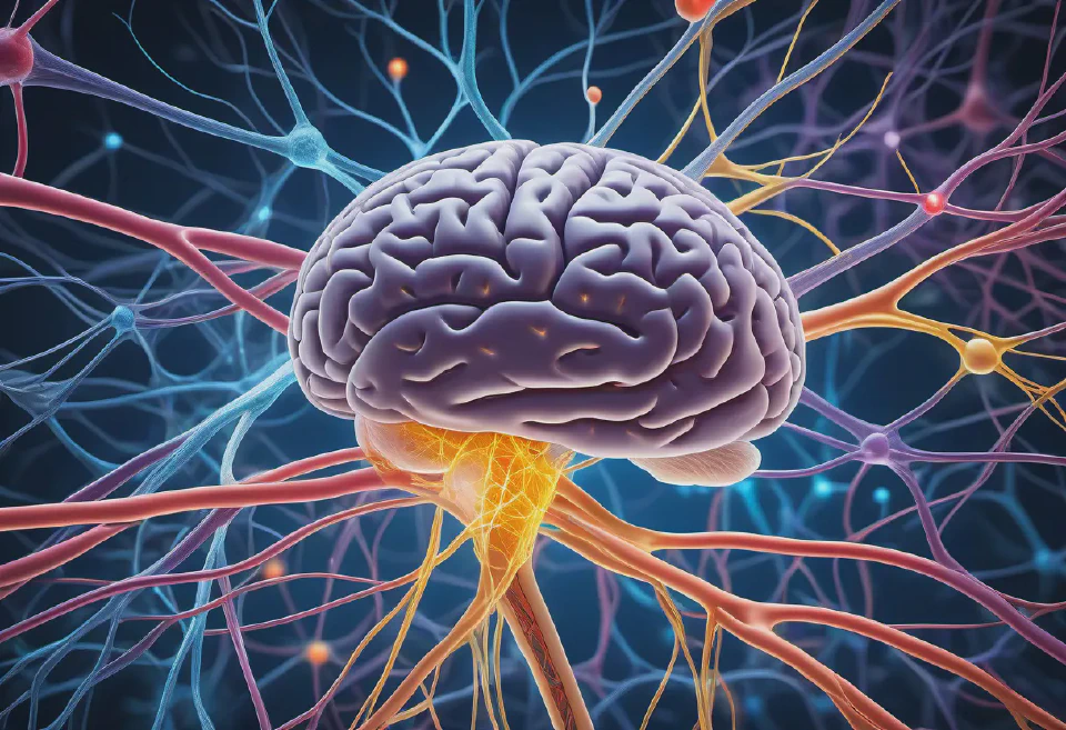
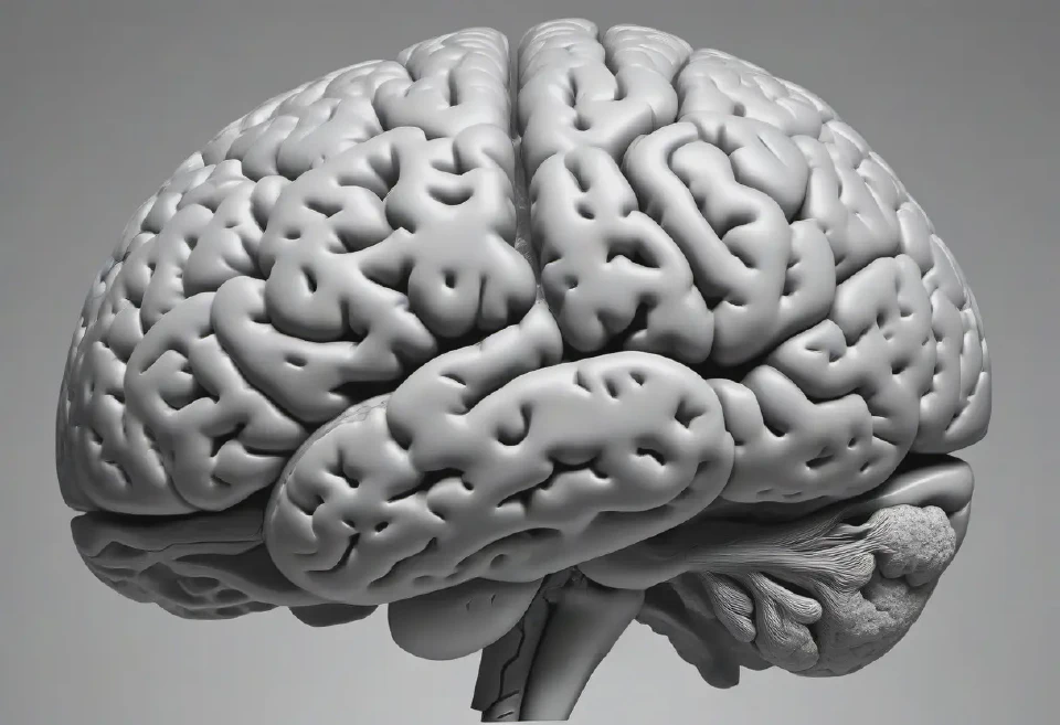
A high-definition illustration of the brain's neural network, featuring detailed depictions of neurons, synapses, and neurotransmitters, designed to aid in explaining neurological conditions and treatments to patients during neurology telemedicine consultations.
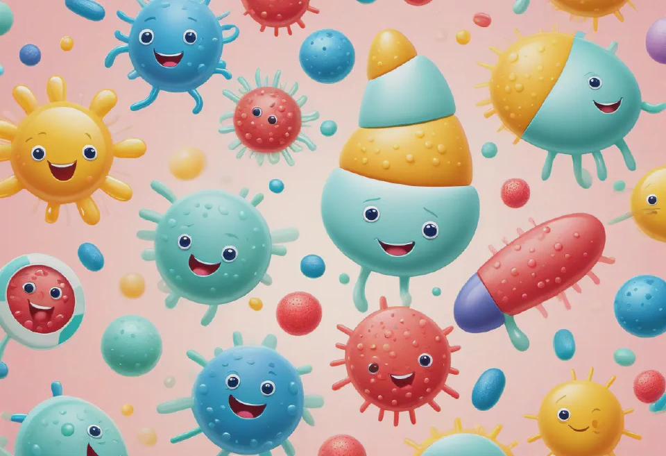
An illustration geared towards pediatric medicines, featuring playful, cartoon-style drawings of friendly bacteria and immune cells. Bright, primary colors against a pastel background should be used to make the packaging appealing to children, incorporating educational elements subtly.
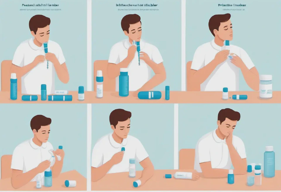
A series of illustrations depicting the correct way to use an inhaler for asthma treatment, including preparation, inhalation, and post-use steps, with each step clearly labeled and depicted in a simple, straightforward style, using soothing colors.
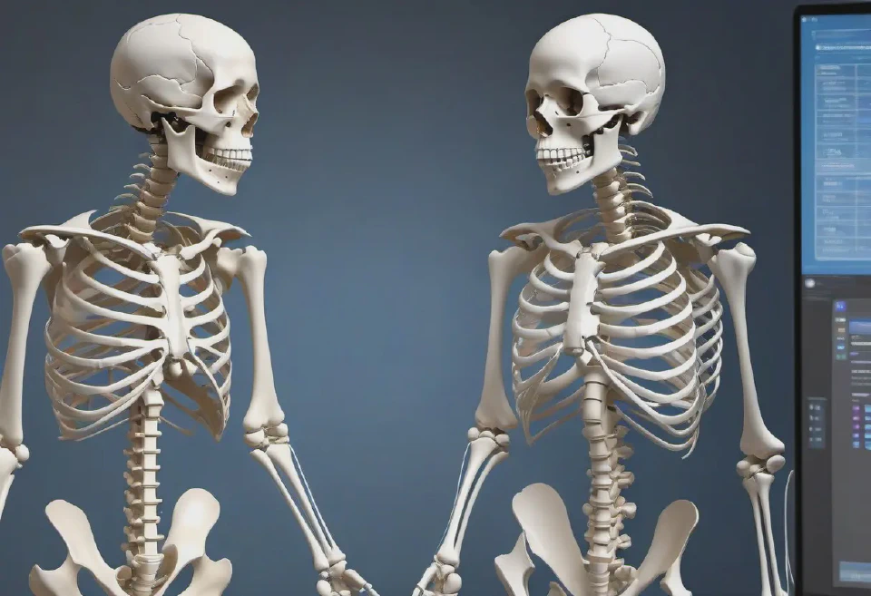
Virtual 3D models of the human skeletal system, highlighting areas affected by different types of arthritis, with the ability to zoom and rotate for detailed inspection during telemedicine sessions.
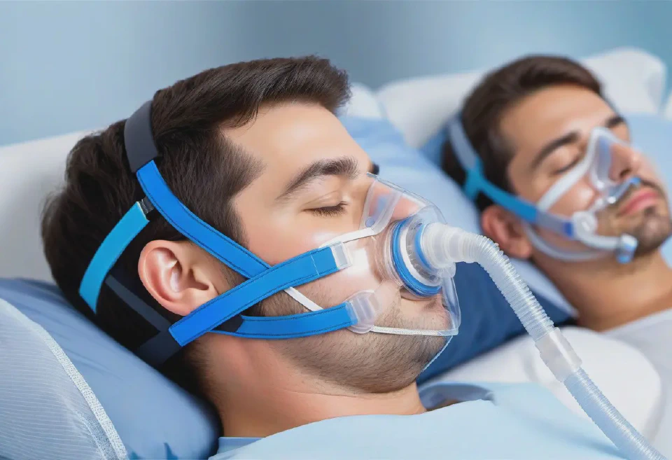
A detailed illustration of setting up and using a continuous positive airway pressure (CPAP) machine for sleep apnea treatment, including the adjustment of straps and mask positioning.
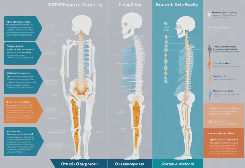
A critical infographic illustrating the progression of osteoporosis, showing normal bone density through to advanced bone thinning and fragility. Use detailed, side-by-side comparisons of bone structure at various stages, highlighting areas prone to fracture and including data on the impact of treatments and lifestyle changes.
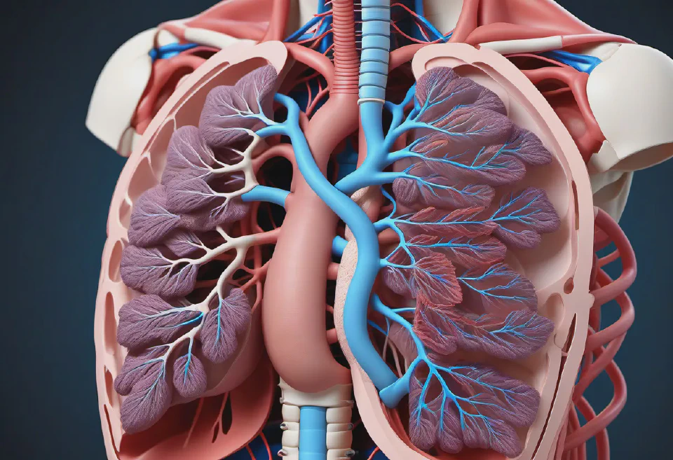
An anatomically detailed 3D model of the human respiratory system, highlighting the trachea, bronchi, and lungs, with sections that can be manipulated to simulate the effects of surgeries like lobectomy or tracheal resection.

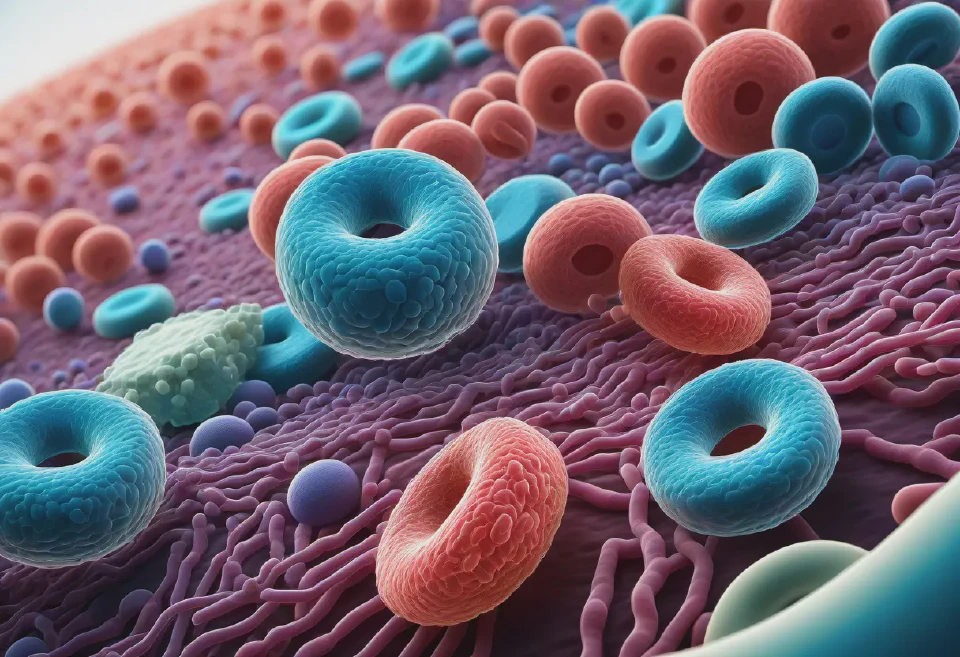
A vivid, high-resolution image of various blood cells, including red blood cells, white blood cells, and platelets, in a dynamic flow within a blood vessel, demonstrating the complexities of the human circulatory system, with a macroscopic lens view.

An illustration of a friendly doctor explaining the process of a knee replacement surgery to a patient using a 3D model of the knee, with welcoming and calming clinic background, vibrant but soft color palette.
A high-resolution, grayscale 3D image of a human brain from an MRI scan, showing detailed structures including the cerebral cortex, ventricles, and cerebellum, with subtle signs of a small, benign tumor in the right hemisphere. Capture in axial view.
8. An infographic summarizing key healthcare statistics such as average life expectancy, common diseases, and healthcare access levels across different demographic groups, using vibrant colors and symbols for clarity.
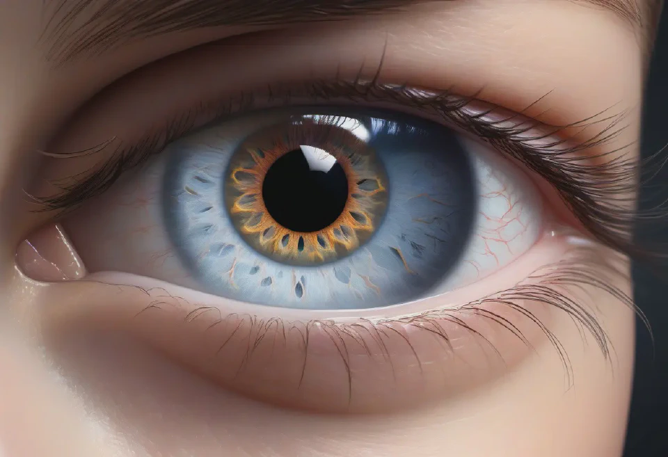
A hyper-realistic rendering of the human eye, showcasing the anatomy in exquisite detail including the iris, cornea, and lens, with annotations for educational use, set against a shadowed background to draw focus to the structures.
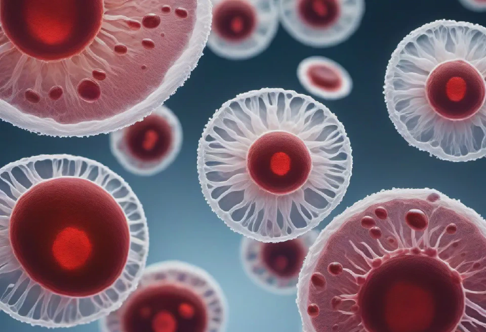
A detailed microscopic image of a blood smear showing various blood cells, including red blood cells, white blood cells, and platelets, with a high level of detail and clarity, utilizing quality modifiers for a realistic look suitable for medical research.
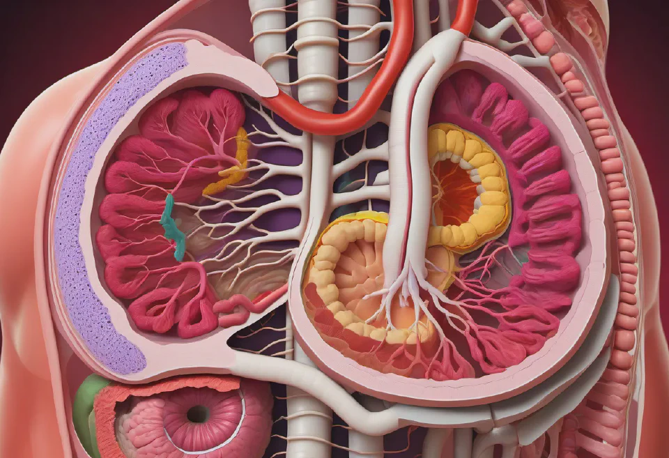
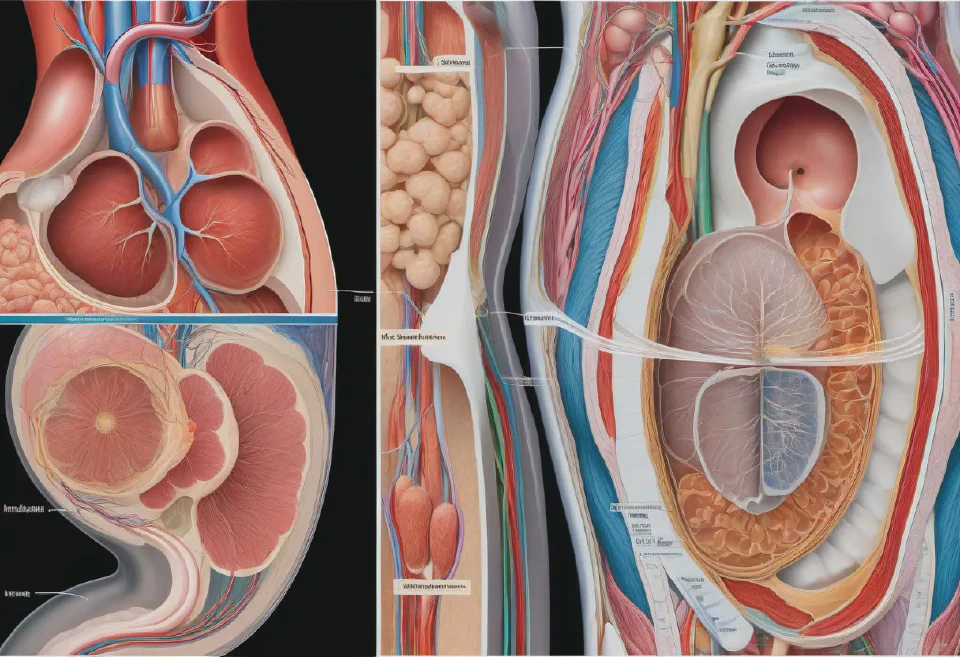
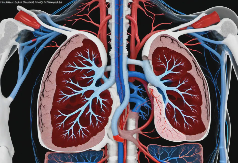
A comprehensive 3D model of the human digestive system, with emphasis on the stomach and intestines, showcasing areas affected by diseases like Crohn's or colorectal cancer for surgical planning.
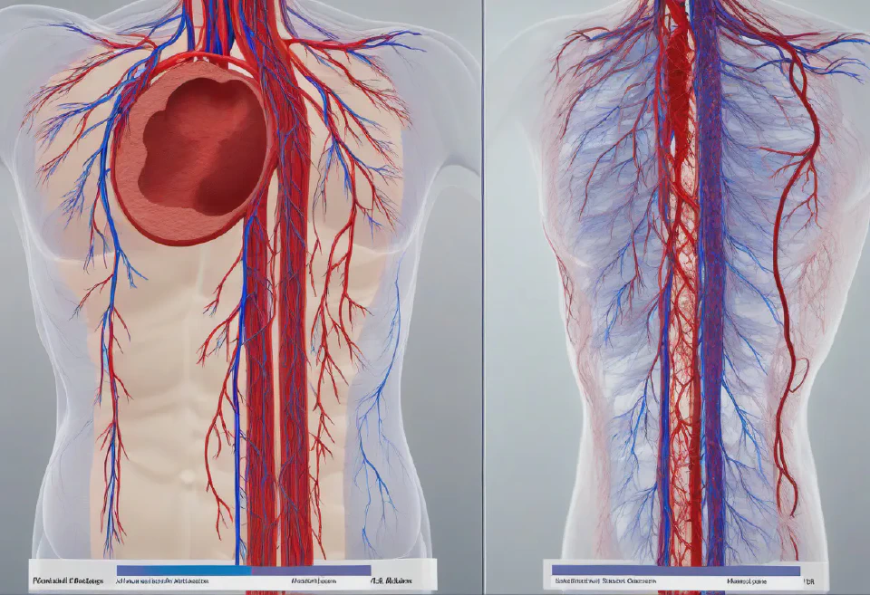
Animated blood flow simulations through various types of blood vessels, demonstrating normal vs. abnormal conditions like blockages or aneurysms, for cardiovascular discussions.

A sleek, precision-engineered insulin pen for diabetes management, showcased on a clean, medical blue background with abstract glucose molecule graphics subtly integrated. High-resolution, 3D rendering, close-up shot.
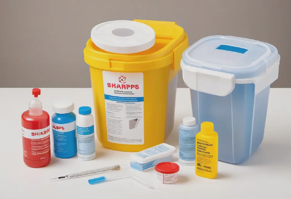
An illustrative step-by-step guide for the safe disposal of sharps used in a medical setting, including syringes and lancets, in a designated sharps disposal container.
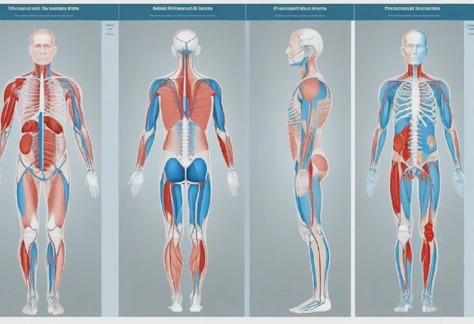
A comprehensive and interactive 3D model of the human muscular system, depicting major muscle groups including the deltoids, biceps, triceps, quadriceps, and gluteus muscles, with detailed textures and colors to reflect muscle types and functions, including animation sequences to illustrate muscle contractions, under dynamic lighting that casts realistic shadows and highlights.
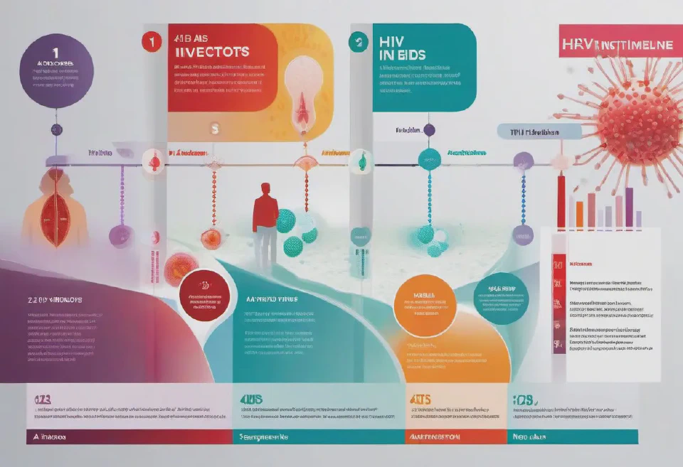
A visual timeline depicting the progression of HIV infection to AIDS, showcasing key stages including acute infection, clinical latency, and AIDS, complete with illustrations of the virus at each stage, its effect on the immune system, and close-ups on CD4 cell count and viral load indicators.
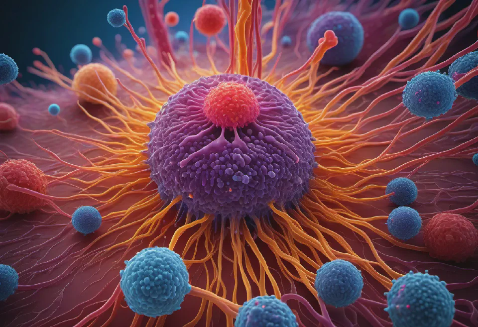
A meticulously crafted, 3D visualization of the human immune response showing a macrophage engulfing a bacterium, with emphasis on the cell interaction, in vibrant colors to detail the process vividly, from a microscopic perspective.

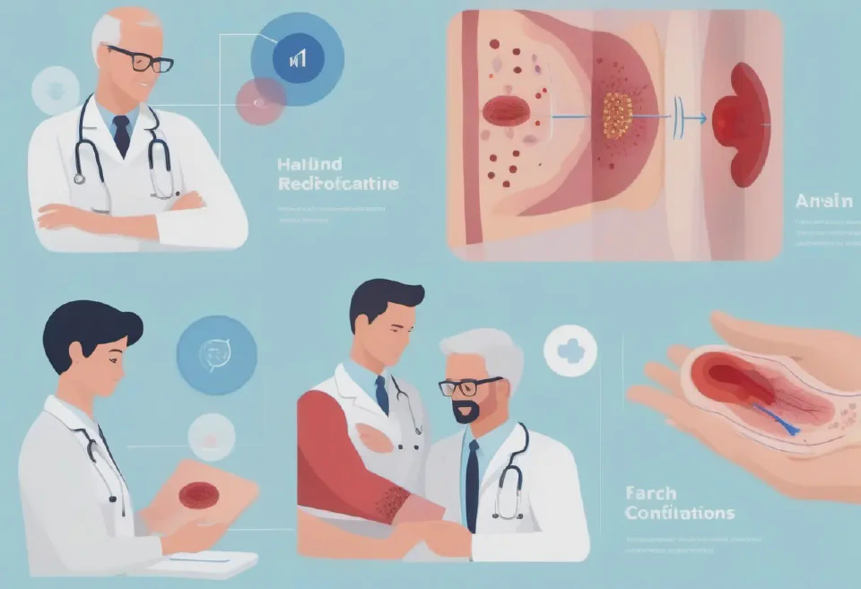
A high-quality, detailed illustration of a healthcare professional demonstrating the correct way to attach a blood pressure cuff on a patient’s arm, ensuring correct positioning and alignment. Include a clear view from a side angle to show how the cuff wraps around the upper arm.
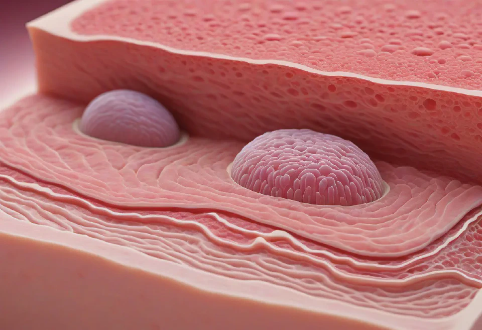
A 3D rendered model of the human skin, displaying its three layers (epidermis, dermis, subcutis) with microscopic accuracy, including hair follicles and sweat glands, and the option to experience the skin's reaction to different environmental conditions.

An outdoor adventure store display themed around camping under the stars. Use a tent set up with cozy sleeping bags, a faux campfire, and atmospheric forest sounds. Highlight the latest in camping technology with QR codes for product details.
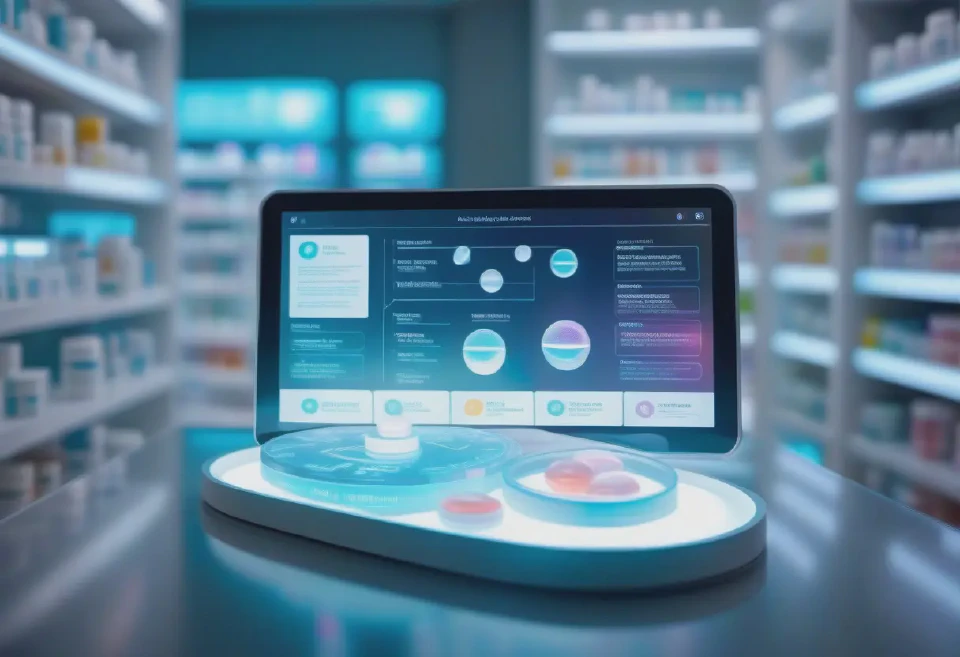
A digital, holographic display of various pharmaceutical pills floating above a smart tablet in a pharmacy setting, showcasing a futuristic medication selection process. High-resolution, medium shot with a focus on the hologram and soft-focus on the background.
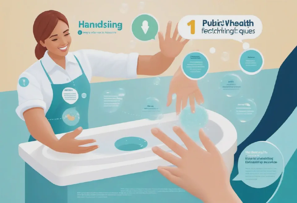
An interactive digital illustration showing a step-by-step guide to proper handwashing techniques, with animated bubbles and hands for a public health brochure.
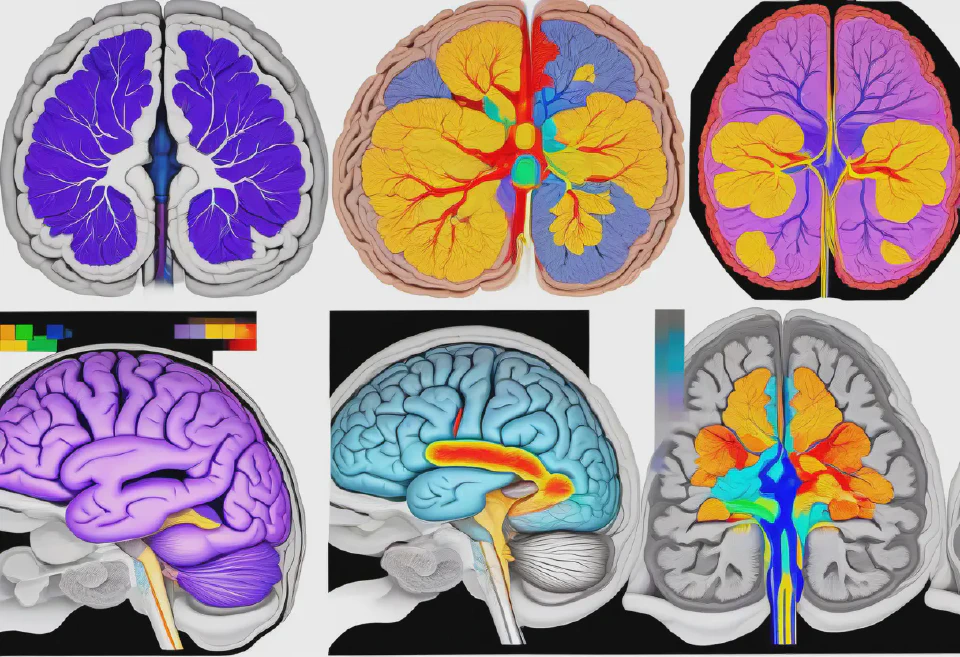
A series of images depicting the progression of Alzheimer's disease in the brain, from early to advanced stages. Each image should show the brain from a top-down view, with color coding to indicate areas of healthy brain tissue, mild cognitive impairment, and severe degeneration.
Interactive diagrams of the human body highlighting areas affected by common postural issues, with sliders to show effects of corrections.
A medical infographic comparing healthy versus diseased lung tissue, including side-by-side images with a high degree of detail and clarity. One side should represent a healthy lung with clear airways, while the other shows a lung affected by chronic obstructive pulmonary disease (COPD), with inflamed airways and mucus build-up.
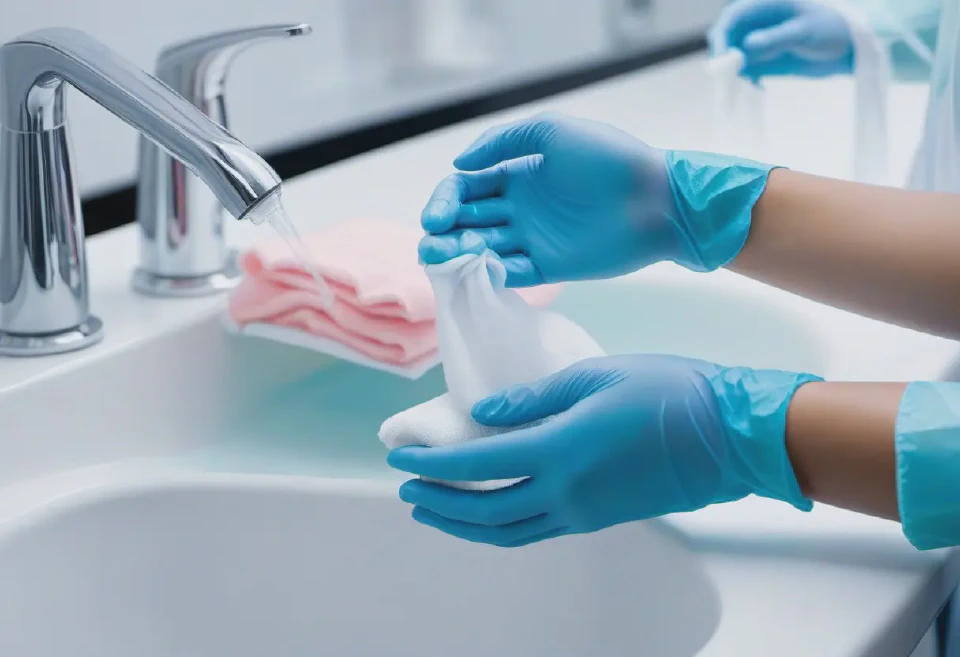
A comprehensive illustration showing the proper method for hand-washing and glove application in a medical environment, emphasizing critical areas of the hands that require thorough cleaning and the correct technique for donning sterile gloves.
An animated sequence depicting the process of wound healing over time, from injury to full recovery, to educate patients during remote consultations.
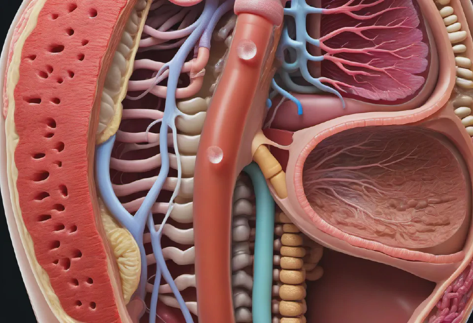
An immersive model of the human digestive system, from the mouth to the anus, highlighting key organs such as the stomach, liver, pancreas, and intestines, with texture-rich rendering for realism, interactive elements to demonstrate the digestion process, and clear, visible labeling for educational purposes, lit with diffuse lighting to emphasize the details and structures.
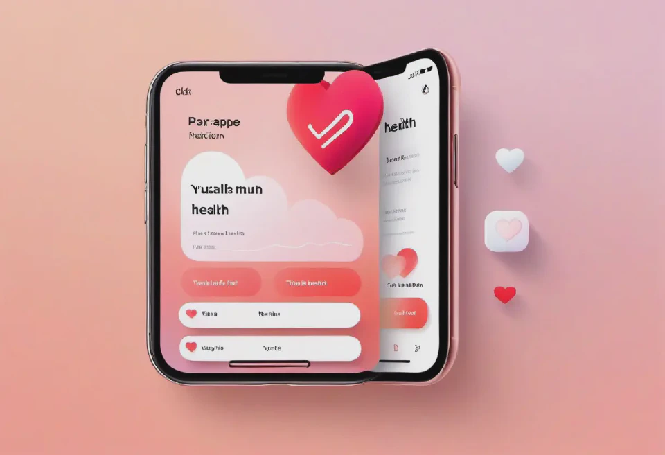
10. A notification pop-up from a health app, illustrated with a heart icon, reminding users to take a moment to breathe and focus on their heart health, set against a soft, gradient background.
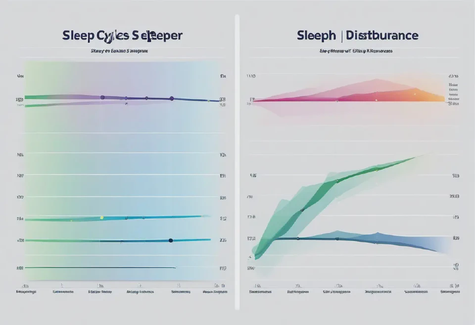
10. An illustrative chart comparing the sleep cycles of a healthy sleeper versus someone with sleep disturbances, using soft, contrasting colors to differentiate between the two. Eye-friendly lighting and a clear, concise layout make the information easily digestible.
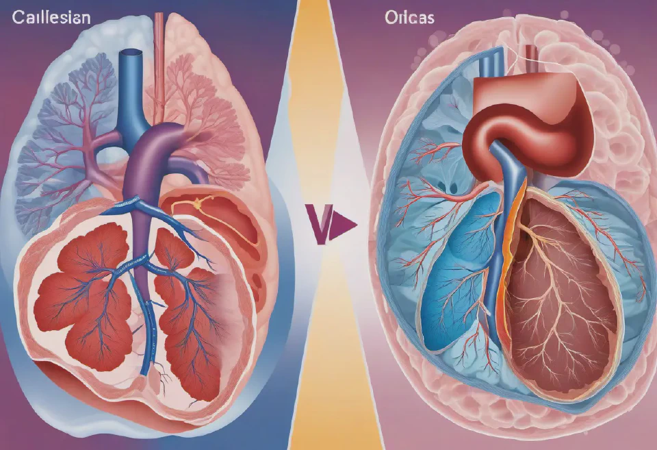
A high-resolution 3D model of a human heart with detailed annotations of the valve structures, viewed in a virtual reality setup for immersive surgical planning.
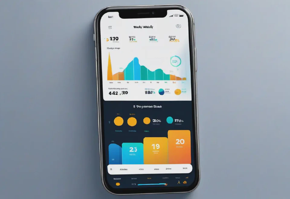
8. An engaging, animated chart within a fitness app showing a user's progress towards their weekly exercise goals, with icons representing different activities like cycling and swimming.
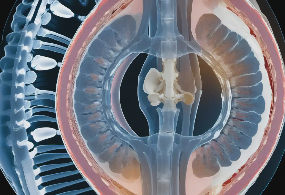
Design a composite MRI image of the entire spinal column, from the cervical to the lumbar regions, showcasing a comprehensive view of the bones, discs, and nerve roots, with an illustrative focus on a herniated disc. Ensure the depiction is in a continuous sagittal view for a holistic educational experience.
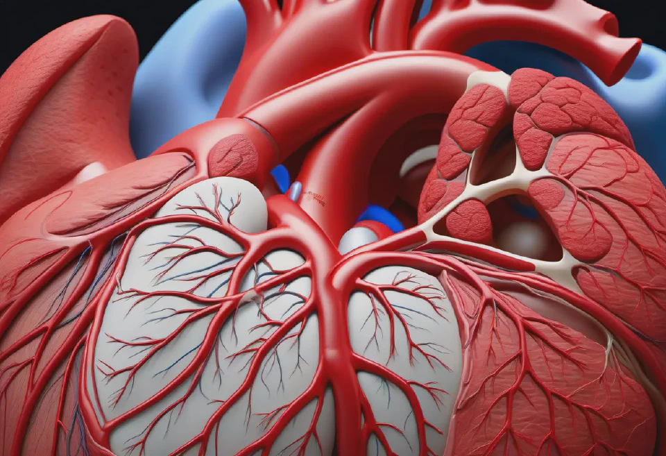
1. A photorealistic 3D rendering of the human heart, showcasing the intricate network of coronary arteries, labeled with high precision in a clear, educational layout for a cardiology conference presentation. The image should have a dynamic lighting setup to emphasize the textures and details of the heart's anatomical structure, captured from an anterior view.
9. A digital journal section in a wellness app, designed with pastel shades and calming imagery of the sky at dusk, encouraging users to reflect on their daily health habits.
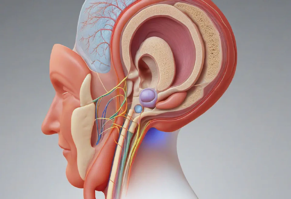
A realistic 3D model of the human ear, including the outer, middle, and inner structures, designed to assist in the planning of surgeries for conditions such as cholesteatoma or otosclerosis.
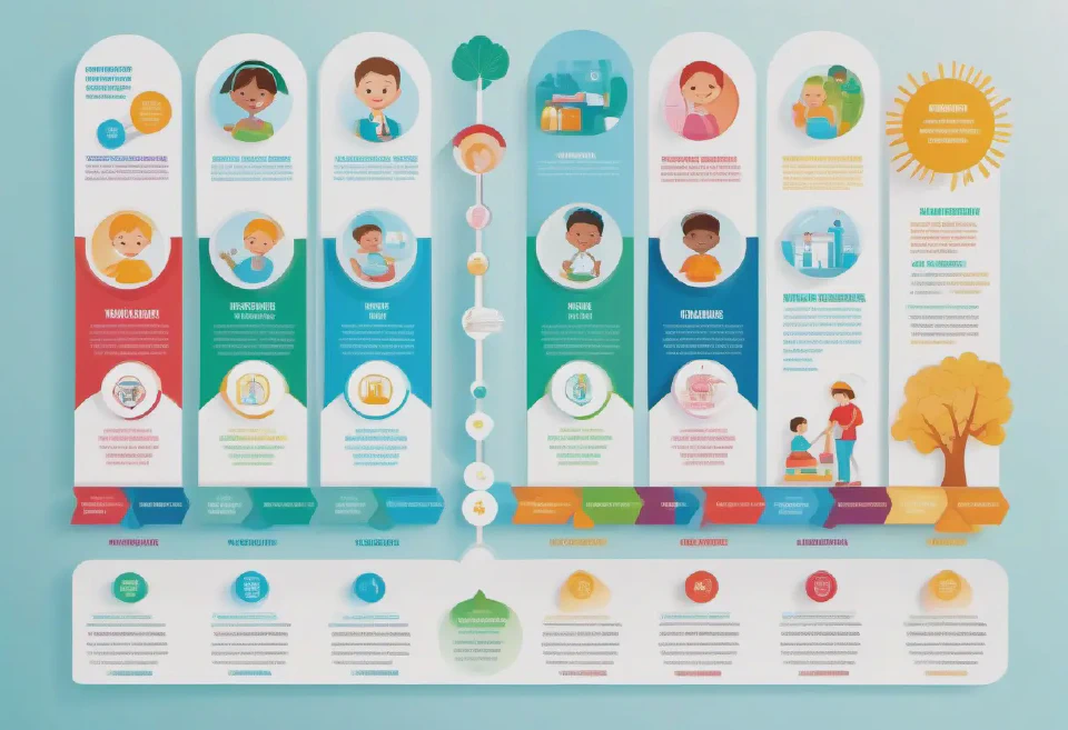
An infographic focused on the benefits of vaccination, presenting a timeline of vaccines a child receives from birth to adolescence, incorporating friendly graphics and icons, executed in a cheerful and colorful style to promote positive messaging.
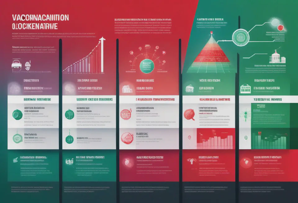
A detailed timeline infographic displaying the evolution of a disease outbreak over months, with icons representing key events (vaccination drives, lockdowns) and a gradient background transitioning from red to green to signify severity to control.
A series of blister packs forming a mosaic of a human brain, illustrating a medication for neurological disorders, positioned on a dark background with neural network-inspired patterns. High-quality, close-up shot, slightly angled perspective.
4. An animated, 3D anatomical model of the human respiratory system responding to an asthma attack, for an online medical education platform. The animation should show the constriction of airways, swelling, and the effect on airflow in high detail, with annotations explaining each process clearly and accurately.

A virtual reality environment simulating the interior of a human ear, to aid in explaining ear infections or hearing loss issues during telehealth sessions.
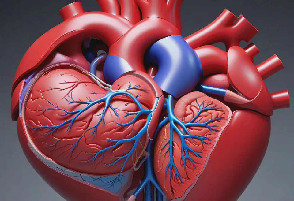
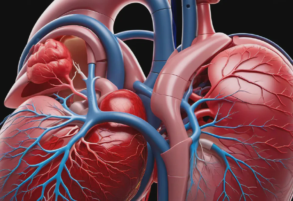
A detailed 3D model of the human heart, showing blood flow and chambers, to explain heart conditions to patients via telemedicine. Include annotations for major parts.

A series of illustrated exercises for carpal tunnel syndrome, including wrist stretches and strengthening activities, presented in a step-by-step format with clear, simple instructions, to support patients in tele-rehabilitation sessions.
10. An image focusing on the human reproductive system, detailing male and female anatomy with separate diagrams that include the internal and external structures, annotated for reproductive biology education.
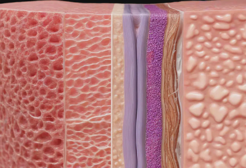
An interactive model of the human integumentary system (skin), showing detailed structures such as the epidermis, dermis, hair follicles, and sweat glands, rendered to mimic skin texture and color variations closely, with zoom and rotation capabilities for a thorough examination, and dynamic, interactive labeling for educational insights into skin health, disease, and care, under uniformly balanced lighting to avoid harsh shadows and highlight textures.
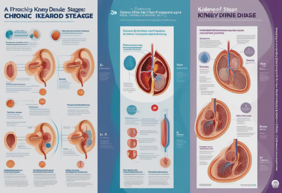
An educational poster series focusing on the progression of chronic kidney disease through its five stages, from slight damage with normal or high filtration to kidney failure. Each poster should depict kidneys at each stage with damage highlighted and key symptoms and potential treatments listed.
An interactive scatter plot representing patient satisfaction scores versus treatment duration, featuring vibrant color coding for different age groups, with a semi-transparent hover detail box for in-depth information.
A highly detailed, scientific illustration for antibiotic packaging, showing a bacterium and the antibiotic mechanism of action at the molecular level. Use accurate, vibrant colors to differentiate between the bacterial structure and the drug, with a magnified, inside-looking-out perspective for dramatic effect.
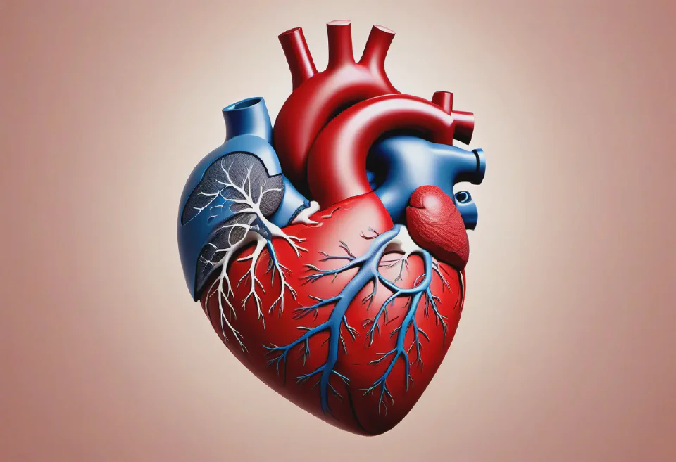
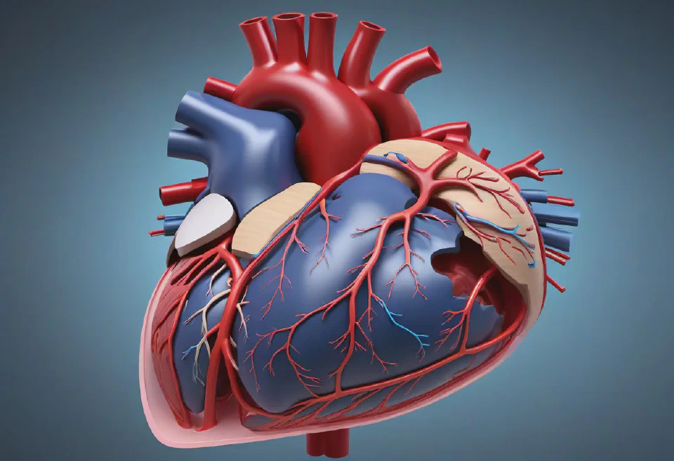
4. A detailed, anatomical illustration of the human heart, with parts labeled to educate users about heart health. The image uses realistic shading and textures to give depth, set against a minimalist background to keep the focus on the educational content.
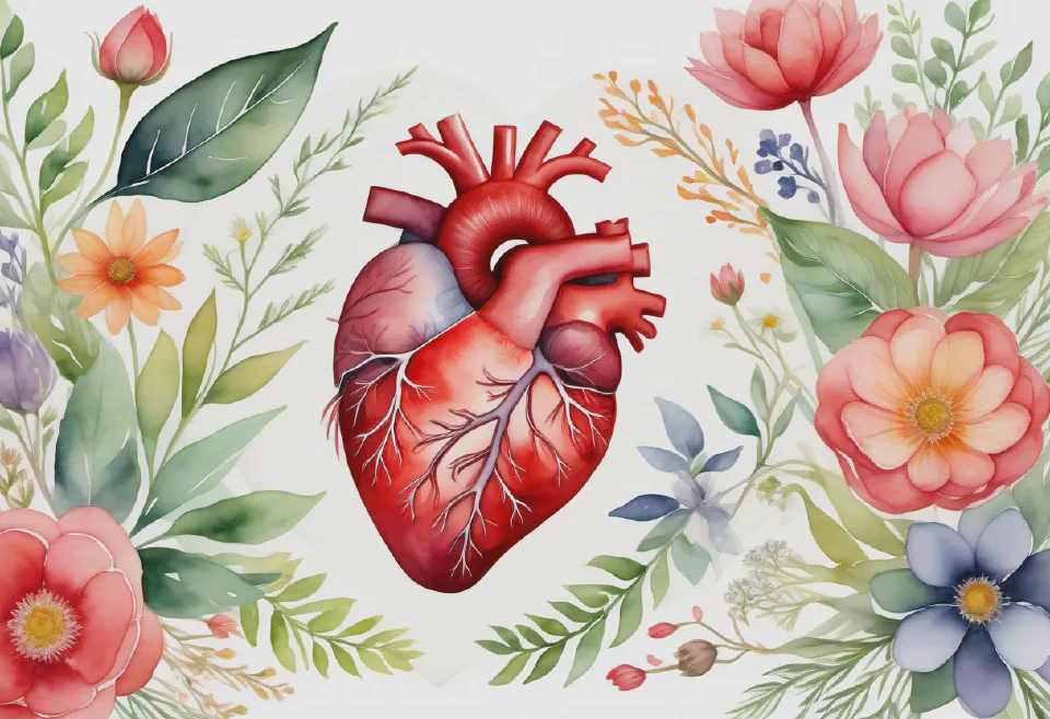
An elegant, watercolor illustration of the human heart connected to various plants and flowers, symbolizing holistic heart health for a cardiovascular health brochure.

A friendly, cartoon-style doctor explaining the food pyramid to children, surrounded by vivid illustrations of healthy foods for a pediatric nutritional guide.
1. A highly detailed virtual reality simulation of a human heart, showing intricate blood vessels and beating in real-time, illuminated from within to emphasize anatomical features, for cardiac surgery training.
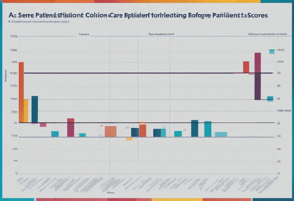
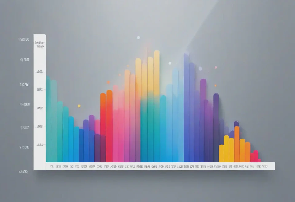
10. A series of histograms comparing patient satisfaction scores before and after implementing a new healthcare policy, with each histogram focusing on a different aspect of patient care, and annotations highlighting significant findings.

3. A dynamic, action-packed image of a diverse group of people jogging in a lush, green park at sunrise, promoting the benefits of regular exercise. The camera angle is low and ahead of the runners, capturing their determination and the beauty of the natural environment.
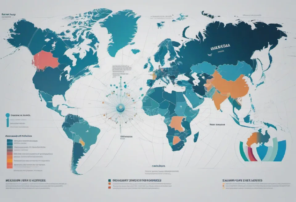
A radar chart comparing the health outcomes of different countries across various metrics (e.g., life expectancy, infant mortality rate), set against a backdrop of a subtly animated, digital world map, highlighting countries as they are discussed.
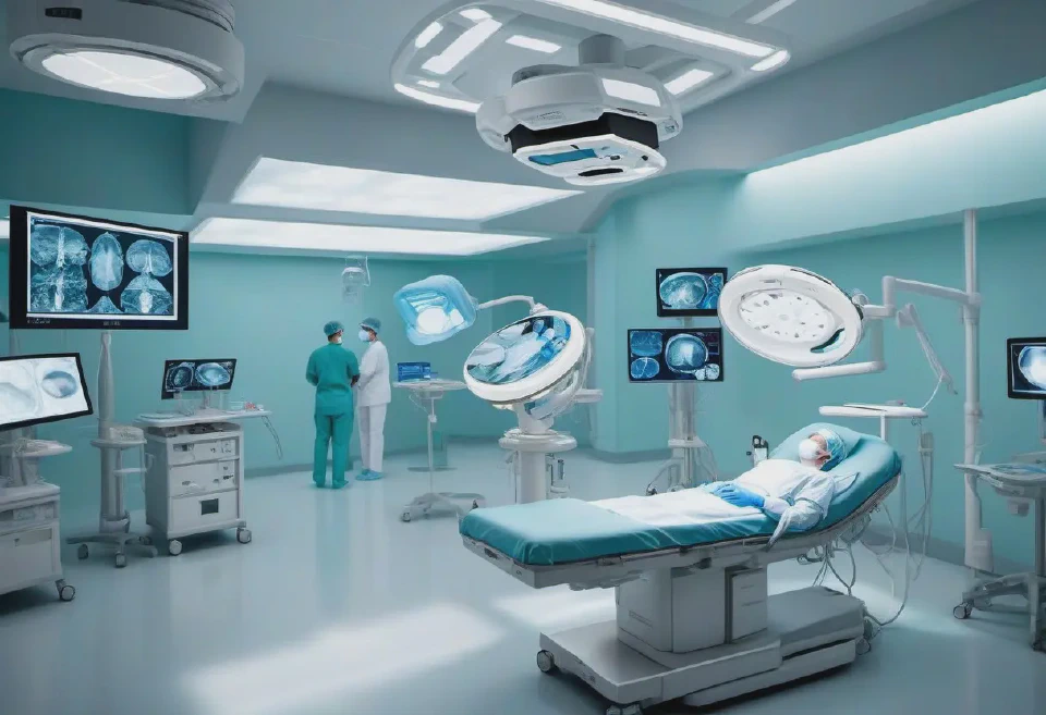
2. An immersive 3D operating room scenario, where medical students can navigate around and interact with equipment and a virtual patient, to practice surgical procedures with high realism under various lighting conditions.

An illustration demonstrating the correct method to apply and remove surgical gloves and masks, highlighting do's and don'ts to ensure sterility.
10. An infographic combining high-quality images and graphs to illustrate the epidemiology of COVID-19, including transmission rates, demographic impacts, vaccine efficacy, and preventive measures for a public health newsletter. The infographic should be visually engaging, using icons, charts, and photos to create a compelling, educational narrative, with a clean layout for easy reading.
1. A high-resolution, 3D scatter plot visualizing patient age vs. treatment effectiveness, with different colors representing various treatments, captured from a bird's-eye view.
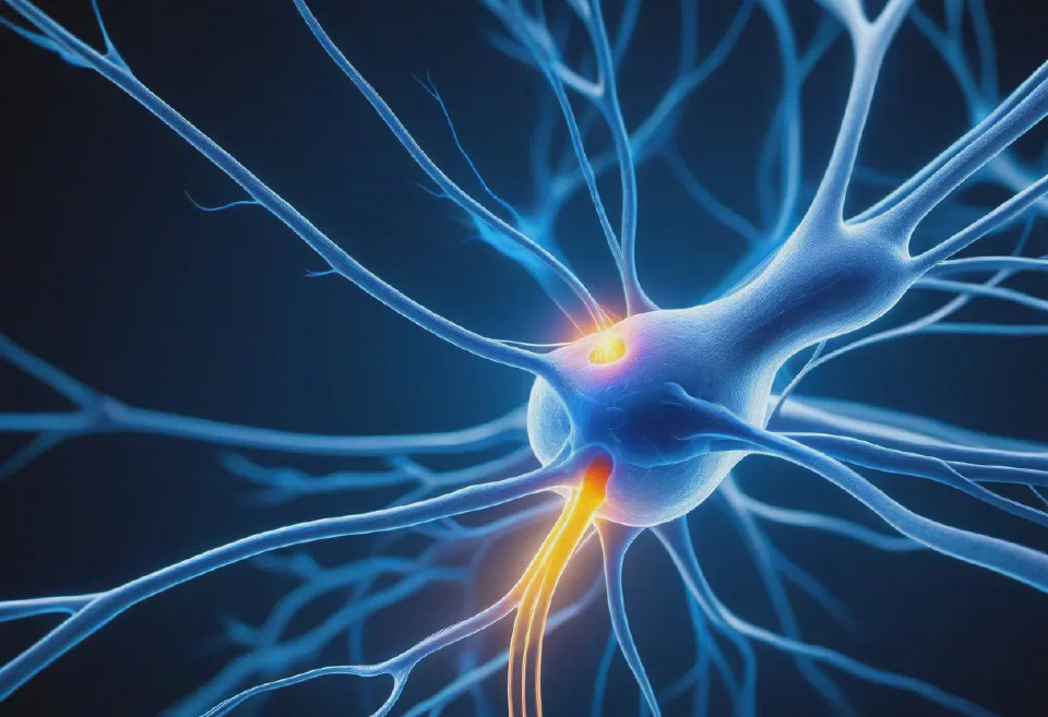
A dynamic, 3D-rendered image of a neuron firing in the human brain, with a focus on the synaptic transmission process. The neuron should glow with electric blue light to indicate activity, against a dark background to enhance visibility.

A set of minimalist geometric patterns for a psychological wellness app's medication reminders, using calming colors like mint green, soft pink, and pale blue. Each medication type could have a unique pattern, combining both aesthetic qualities and functional clarity in a digital format.
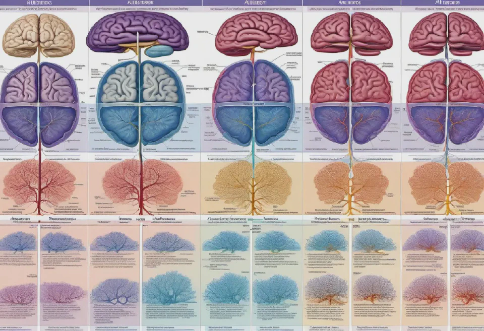
A high-resolution, digitally rendered timeline representing the progression of Alzheimer's disease, including early, moderate, and severe stages, illustrated with detailed brain images showing changes in neural pathways and atrophy areas. The image should have a side-by-side comparison format and be labeled accurately for educational use.
An intricate CT scan image of the thoracic cavity, highlighting the heart, lungs, and major blood vessels with high contrast, simulating an early stage of lung cancer. Viewed in coronal plane with annotations for educational purposes.
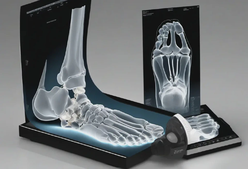
Construct a CT scan of the foot, detailing the bones, joints, and soft tissues with high contrast, including a simulated case of a calcaneal fracture. Desired perspective is in axial view, prioritizing detail to aid in the training of identifying fractures.

2. An interactive heatmap showing the geographic distribution of a specific disease outbreak across a city, with varying shades of red indicating areas of higher density.
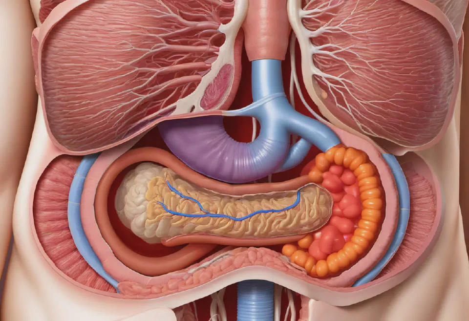
7. An in-depth look at the human digestive system, from the oral cavity to the large intestine, showcasing the digestion and nutrient absorption processes with precise labeling for gastroenterology students.
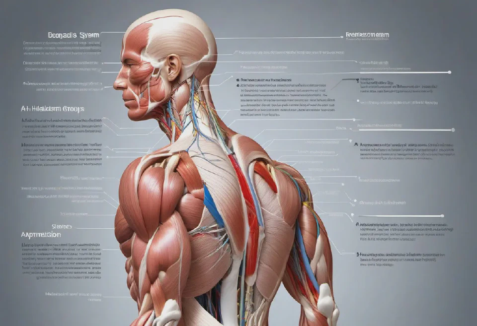
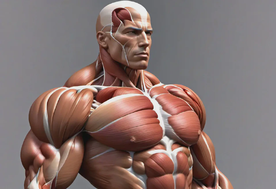
6. A realistic, muscular system illustration capturing the major muscle groups of the human body, including both superficial and deep muscles, annotated with names and functions for physical therapy education.

5. A highly detailed, schematic illustration of the molecular mechanism of a CRISPR-Cas9 gene-editing system targeting a DNA strand, intended for the cover of a genomic research publication. The image should use vibrant colors to differentiate components and include dynamic, educational annotations detailing each step of the editing process.